 E-submission
Search
- Page Path
- HOME > Search
Review Article
- Contrast-enhanced Ultrasonography: The Third Modality for Differentiation of Liver Mass
- Min Kyu Kang, Moon Young Kim, Seong Hee Kang, Soon Koo Baik
- J Liver Cancer. 2019;19(2):91-96. Published online September 30, 2019
- DOI: https://doi.org/10.17998/jlc.19.2.91
- 4,657 Views
- 96 Downloads
- 1 Citation
-
 Abstract
 PDF
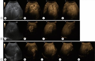
PDF - Contrast-enhanced ultrasonography (CEUS) using microbubble ultrasonography contrast agent can show the vascular structure and unique contrast enhancement patterns of focal liver lesions, including hepatocellular carcinoma (HCC). CEUS shows three phases, similar to a vascular pattern on computer tomography (CT), and typical arterial enhancement and portal or late phase washout in HCC. CEUS can show real-time images without nephrotoxicity or radiation hazard and can be used as guidance for loco-regional treatment and estimation of treatment response of HCC. In addition, some data recently revealed the usefulness of CEUS in the early estimation of response to anti-cancer pharmacological (i.e., sorafenib) therapy in advanced HCC. Although CEUS has limitations in clinical practice and more investigation is needed for its validation, it is recommended as a main diagnostic modality in a few major clinical practice guidelines for HCC. Thus, greater understanding of CEUS is necessary to extend its application in real practice for diagnosis and management of diseases.
-
Citations
Citations to this article as recorded by
- Perfluorobutane-Enhanced Ultrasound for Characterization of Hepatocellular Carcinoma From Non-hepatocellular Malignancies or Benignancy: Comparison of Imaging Acquisition Methods
Seungchul Han, Se Woo Kim, Sungeun Park, Jeong Hee Yoon, Hyo-Jin Kang, Jeongin Yoo, Ijin Joo, Jae Seok Bae, Jeong Min Lee
Ultrasound in Medicine & Biology.2023; 49(10): 2256. CrossRef
- Perfluorobutane-Enhanced Ultrasound for Characterization of Hepatocellular Carcinoma From Non-hepatocellular Malignancies or Benignancy: Comparison of Imaging Acquisition Methods

Original Article
- Follow-up of Hepatocellular Carcinoma After Transarterial Chemoembolization; The Concordance of Contrast Enhanced Ultrasonography and Lipiodol CT
- Gene Hyun Bok, Soung Won Jeong, Jae Young Jang, Sae Hwan Lee, Sang Gyune Kim, Sang-Woo Cha, Young Seok Kim, Young Deok Cho, Hong Soo Kim, Boo Sung Kim
- J Liver Cancer. 2014;14(2):115-119. Published online September 30, 2014
- DOI: https://doi.org/10.17998/jlc.14.2.115
- 889 Views
- 4 Downloads
-
Abstract
PDF
- Background/Aim
s: The aim of this study is to evaluate the concordance of contrast-enhanced ultrasonography (CEUS) and lipiodol computed tomography (L-CT) for the assessment of viable hepatocellular carcinoma (HCC) after transarterial chemoembolization (TACE).
Methods
We retrospectively reviewed the post-TACE CEUS and L-CT images of 65 consecutive HCCs in 41 patients to assess the presence of viable tumor tissue. Forty-seven HCCs in 31 patients that underwent post-TACE L-CT within 4 weeks of the CEUS examination were included. The degree of concordance between CEUS and L-CT and factors related to concordance were analyzed.
Results
The overall concordance of CEUS and LDCT was 78.7% (37/47). The concordance with L-CT for viable tumor and non-viable tumor tissue on CEUS was 95.2%, and 65.4% respectively (P<0.013). Diffuse tumors had a tendency for non-concordance (P=0.066). Although 3 of 4 lesions located in the hepatic dome were non-concordant, the sample size was too small to establish significance. The mean tumor size for concordant and non-concordant tumors was 2.9 and 3.0 cm, with no significant difference.
Conclusions
Although the concordance of CEUS and L-CT for viable tumor tissue was high, the concordance for non-viable tumor tissue was relatively low. Prospective studies using angiography as a gold standard should be performed in the future. (J Liver Cancer 2014;14:115-119)
 First
First Prev
Prev


 Follow JLC on Twitter