 E-submission
Search
- Page Path
- HOME > Search
Original Article
- Additional nodules detected using EOB-MRI in patients with resectable single hepatocellular carcinoma: an implication for active treatment strategy
- Na Reum Kim, Seoung Yoon Rho, Jonathan Navarro, Chansik An, Dai Hoon Han, Jin Sub Choi, Myeong-Jin Kim, Gi Hong Choi
- J Liver Cancer. 2024;24(1):92-101. Published online February 14, 2024
- DOI: https://doi.org/10.17998/jlc.2024.01.25
- 912 Views
- 46 Downloads
-
 Abstract
 PDF
PDF - Background/Aim
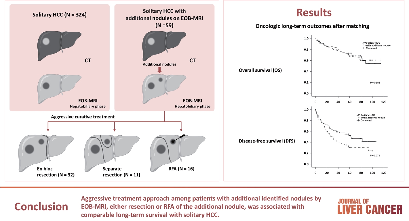
Gadolinium-ethoxybenzyl-diethylenetriamine pentaacetic acid-enhanced magnetic resonance imaging (EOBMRI) further enhances the identification of additional hepatic nodules compared with computed tomography (CT) alone; however, the optimal treatment for such additional nodules remains unclear. We investigated the long-term oncological effect of aggressive treatment strategies for additional lesions identified using EOB-MRI in patients with hepatocellular carcinoma (HCC).
Methods
Data from 522 patients diagnosed with solitary HCC using CT between January 2008 and December 2012 were retrospectively reviewed. Propensity score-matched (PSM) analysis was used to compare the oncologic outcomes between patients with solitary HCC and those with additional nodules on EOB-MRI after aggressive treatment (resection or radiofrequency ablation [RFA]).
Results
Among the 383 patients included, 59 had additional nodules identified using EOB-MRI. Compared with patients with solitary HCC, those with additional nodules on EOB-MRI had elevated total bilirubin, aspartate transaminase, and alanine transaminase; had a lower platelet count, higher MELD score, and highly associated with liver cirrhosis (P<0.05). Regarding long-term outcomes, 59 patients with solitary HCC and those with additional nodules after PSM were compared. Disease-free survival (DFS) and overall survival (OS) were comparable between the two groups (DFS, 60.4 vs. 44.3 months, P=0.071; OS, 82.8 vs. 84.8 months, P=0.986).
Conclusion
The aggressive treatment approach, either resection or RFA, for patients with additional nodules identified on EOBMRI was associated with long-term survival comparable with that for solitary HCC. However, further studies are required to confirm these findings.

Review Articles
- Radiologic features of hepatocellular carcinoma related to prognosis
- Shin Hye Hwang, Hyungjin Rhee
- J Liver Cancer. 2023;23(1):143-156. Published online March 9, 2023
- DOI: https://doi.org/10.17998/jlc.2023.02.16

- 2,089 Views
- 113 Downloads
- 2 Citations
-
Abstract
PDF
- The cross-sectional imaging findings play a crucial role in the diagnosis of hepatocellular carcinoma (HCC). Recent studies have shown that imaging findings of HCC are not only relevant for the diagnosis of HCC, but also for identifying genetic and pathologic characteristics and determining prognosis. Imaging findings such as rim arterial phase hyperenhancement, arterial phase peritumoral hyperenhancement, hepatobiliary phase peritumoral hypointensity, non-smooth tumor margin, low apparent diffusion coefficient, and the LR-M category of the Liver Imaging-Reporting and Data System have been reported to be associated with poor prognosis. In contrast, imaging findings such as enhancing capsule appearance, hepatobiliary phase hyperintensity, and fat in mass have been reported to be associated with a favorable prognosis. Most of these imaging findings were examined in retrospective, single-center studies that were not adequately validated. However, the imaging findings can be applied for deciding the treatment strategy for HCC, if their significance can be confirmed by a large multicenter study. In this literature, we would like to review imaging findings related to the prognosis of HCC as well as their associated clinicopathological characteristics.
-
Citations
Citations to this article as recorded by
- Radiomics and machine learning based on preoperative MRI for predicting extrahepatic metastasis in hepatocellular carcinoma patients treated with transarterial chemoembolization
Gang Peng, Xiaojing Cao, Xiaoyu Huang, Xiang Zhou
European Journal of Radiology Open.2024; 12: 100551. CrossRef - Imaging prognostication and tumor biology in hepatocellular carcinoma
Diana Kadi, Marilyn F. Yamamoto, Emily C. Lerner, Hanyu Jiang, Kathryn J. Fowler, Mustafa R. Bashir
Journal of Liver Cancer.2023; 23(2): 284. CrossRef
- Radiomics and machine learning based on preoperative MRI for predicting extrahepatic metastasis in hepatocellular carcinoma patients treated with transarterial chemoembolization
- Conventional Chemoembolization for Hepatocellular Carcinoma: Role of Cone-Beam Computed Tomography Guidance
- In Joon Lee, Jin Wook Chung
- J Liver Cancer. 2019;19(1):19-29. Published online March 31, 2019
- DOI: https://doi.org/10.17998/jlc.19.1.19
- 5,288 Views
- 146 Downloads
- 4 Citations
-
Abstract
PDF
- Conventional chemoembolization using Lipiodol-based regimens was introduced in the 1980s, and it is currently recommended as the primary treatment modality for patients with unresectable, intermediate, or locally advanced hepatocellular carcinoma (HCC) by the international guidelines. For better therapeutic efficacy and safety, chemoembolization should be performed as selectively as possible through tumor-feeding arteries, based on the detection of arterial supply to the HCC. With the technical advancement of flat-panel detector, cone-beam computed tomography (CBCT) is mounted on the C-arm of the angiographic machine. CBCT facilitates the detection of small occult HCCs and fine tumor-feeding arteries, recognition of extrahepatic collateral supply, navigation of a microcatheter to the target feeding arteries, prevention of non-target embolization, and intraprocedural assessment of the completeness of treatment with chemoembolization. These functions performed by CBCT ultimately improve the safety and efficacy of chemoembolization and may contribute to improving the prognosis of the patient with HCC.
-
Citations
Citations to this article as recorded by- Transarterial Chemoembolization for Hepatocellular Carcinoma: 2023 Expert Consensus-Based Practical Recommendations of the Korean Liver Cancer Association
Yuri Cho, Jin Woo Choi, Hoon Kwon, Kun Yung Kim, Byung Chan Lee, Hee Ho Chu, Dong Hyeon Lee, Han Ah Lee, Gyoung Min Kim, Jung Suk Oh, Dongho Hyun, In Joon Lee, Hyunchul Rhim
Korean Journal of Radiology.2023; 24(7): 606. CrossRef - Transarterial chemoembolization for hepatocellular carcinoma: 2023 expert consensus-based practical recommendations of the Korean Liver Cancer Association
Yuri Cho, Jin Woo Choi, Hoon Kwon, Kun Yung Kim, Byung Chan Lee, Hee Ho Chu, Dong Hyeon Lee, Han Ah Lee, Gyoung Min Kim, Jung Suk Oh, Dongho Hyun, In Joon Lee, Hyunchul Rhim
Journal of Liver Cancer.2023; 23(2): 241. CrossRef - Transarterial chemoembolization for hepatocellular carcinoma: 2023 Expert consensus-based practical recommendations of the Korean Liver Cancer Association
Yuri Cho, Jin Woo Choi, Hoon Kwon, Kun Yung Kim, Byung Chan Lee, Hee Ho Chu, Dong Hyeon Lee, Han Ah Lee, Gyoung Min Kim, Jung Suk Oh, Dongho Hyun, In Joon Lee, Hyunchul Rhim
Clinical and Molecular Hepatology.2023; 29(3): 521. CrossRef - The efficacy of TACE; how can automated feeder software help?
Hassan Abdelsalam, Doaa M. Emara, Ehab M. Hassouna
Egyptian Journal of Radiology and Nuclear Medicine.2022;[Epub] CrossRef
- Transarterial Chemoembolization for Hepatocellular Carcinoma: 2023 Expert Consensus-Based Practical Recommendations of the Korean Liver Cancer Association
Original Articles
- Transarterial Chemoembolization versus Radiofrequency Ablation for Small Hepatocellular Carcinomas with Discrepant Features on Computed Tomography and Magnetic Resonance Imaging
- Young Youn Cho, Jung Hee Kwon, Jeong-Hoon Lee, Jeong Min Lee, Jae Young Lee, Hyo-Choel Kim, Jin Wook Chung, Won-mook Choi, Eun Ju Cho, Yoon Jun Kim, Jung-Hwan Yoon, Chung Yong Kim, Hyo-Suk Lee
- J Liver Cancer. 2015;15(1):19-29. Published online March 31, 2015
- DOI: https://doi.org/10.17998/jlc.15.1.19
- 1,239 Views
- 8 Downloads
-
Abstract
PDF
- Background/Aims
This study compared the outcomes of patients with small hepatocellular carcinomas (HCCs) who were treated using transarterial chemoembolization (TACE) or radiofrequency ablation (RFA).
Methods
This was a post-hoc analysis of a prospective study that evaluated the diagnostic efficacy of magnetic resonance imaging (MRI) and computed tomography (CT). We analyzed 41 small hepatic nodules in 32 patients that showed typical radiologic hallmarks on both CT and gadoxate-enhanced MRI (typical nodules) and 25 small hepatic nodules from 22 patients that showed atypical radiologic hallmarks on CT and typical radiologic hallmarks on MRI (discrepant nodules).
Results
There were no significant differences in the baseline characteristics of the patients with typical and discrepant nodules. Complete response rates 1 month after TACE or RFA were 75.0% (18/24) and 94.1% (16/17; P=0.20), respectively, for the patients with typical nodules and 58.8% (10/17) and 100% (8/8; P=0.05), respectively, for the patients with discrepant nodules. Treatment failure rates after TACE or RFA were 33.3% (8/24) and 5.8% (1/17; P=0.15), respectively, for the patients with typical nodules and 47.0% (8/17) and 0.0% (0/8; P=0.02), respectively, for the patients with discrepant nodules. Among patients achieving complete response, there were no significant differences in the risk of marginal recurrence.
Conclusions
RFA provided higher complete response rates and significantly lower treatment failure rates than TACE for patients with discrepant nodules of HCC. Therefore, a treatment modality such as RFA may be preferable for small HCCs which show discrepancy on two imaging modalities.
- Follow-up of Hepatocellular Carcinoma After Transarterial Chemoembolization; The Concordance of Contrast Enhanced Ultrasonography and Lipiodol CT
- Gene Hyun Bok, Soung Won Jeong, Jae Young Jang, Sae Hwan Lee, Sang Gyune Kim, Sang-Woo Cha, Young Seok Kim, Young Deok Cho, Hong Soo Kim, Boo Sung Kim
- J Liver Cancer. 2014;14(2):115-119. Published online September 30, 2014
- DOI: https://doi.org/10.17998/jlc.14.2.115
- 904 Views
- 4 Downloads
-
Abstract
PDF
- Background/Aims
The aim of this study is to evaluate the concordance of contrast-enhanced ultrasonography (CEUS) and lipiodol computed tomography (L-CT) for the assessment of viable hepatocellular carcinoma (HCC) after transarterial chemoembolization (TACE).
Methods
We retrospectively reviewed the post-TACE CEUS and L-CT images of 65 consecutive HCCs in 41 patients to assess the presence of viable tumor tissue. Forty-seven HCCs in 31 patients that underwent post-TACE L-CT within 4 weeks of the CEUS examination were included. The degree of concordance between CEUS and L-CT and factors related to concordance were analyzed.
Results
The overall concordance of CEUS and LDCT was 78.7% (37/47). The concordance with L-CT for viable tumor and non-viable tumor tissue on CEUS was 95.2%, and 65.4% respectively (P<0.013). Diffuse tumors had a tendency for non-concordance (P=0.066). Although 3 of 4 lesions located in the hepatic dome were non-concordant, the sample size was too small to establish significance. The mean tumor size for concordant and non-concordant tumors was 2.9 and 3.0 cm, with no significant difference.
Conclusions
Although the concordance of CEUS and L-CT for viable tumor tissue was high, the concordance for non-viable tumor tissue was relatively low. Prospective studies using angiography as a gold standard should be performed in the future. (J Liver Cancer 2014;14:115-119)
Review Articles
- Diagnostic Performance of CT and MRI for Small HCC Less Than 1 cm
- Jung Hoon Kim
- Journal of the Korean Liver Cancer Study Group. 2013;13(2):105-113. Published online September 30, 2013
- DOI: https://doi.org/10.17998/jlc.13.2.105
- 977 Views
- 10 Downloads
-
Abstract
PDF
- Hepatocellular carcinoma (HCC) is the most common primary liver cancer. Worldwide, the surveillance program of HCC is well-established for the patients with high risk. The early stage of HCC has the highest likelihood for cure. It is therefore important to make the early diagnosis of HCC. The imaging criteria for the diagnosis of HCC have established detailing the characteristic enhancement pattern of HCC including hypervascularity in the arterial phase and washout in the portal or delayed phase of dynamic imaging. However, in the current practical guidelines, there are controversies for HCC smaller than 1 cm in size because of the low sensitivity of CT and MR for HCC smaller than 1 cm in size. Recent advances in CT and MR hardware and software allow for rapid acquisition times that can reduce many of motion artifacts. Furthermore, recently introduced hepatocyte-specific MR contrast agents have showed its superior efficacy in both detection and characterization of small focal liver lesion by providing both dynamic imaging and hepatobiliary phase imaging. So, we reviewed the diagnostic performance of CT and MRI for HCC smaller than 1 cm in size. In addition, we introduced our experience for HCC smaller than 1 cm in size.
- Radiology of the Combined HCC and CC
- Jin-Young Choi
- Journal of the Korean Liver Cancer Study Group. 2007;7(1):16-21. Published online June 30, 2007
- 593 Views
- 8 Downloads
-
Abstract
PDF
- Combined hepatocellular-cholangiocarcinoma (HCC-CC) is a rare tumor showing histological evidence of both hepatocellular and biliary epithelial differentiation. Because of its rarity, there have been few reports about the imaging findings of this tumor. The CT findings of the combined HCC-CC include well-defined tumor with signs of malignancy such as hepatic hypervascularity, biliary obstruction, satellite nodules, and lymphadenopathy. Some portions may show hypoattenuation on arterial phase, whereas other portions show delayed persistent enhancement. The signal intensity of this tumor on magnetic resonance (MR) imaging is not specific. However, when the tumor resembles cholangiocarcinoma on gross pathologic exam, it shows peripheral rim-like enhancement, infiltrative margin, and portal vein invasion in patients with liver cirrhosis. On the other hand, when the tumor has gross appearance of HCC, imaging findings are not helpful for diagnosis. Cross-sectional imagings are helpful for predicting the predominant component of the combined HCC-CC and determining the treatment strategy.
Case Reports
- Hepatoid Adenocarcinoma of the Stomach with Liver Metastasis Mimicking Hepatocellular Carcinoma
- Se Hyung Kim, Byung Ihn Choi
- Journal of the Korean Liver Cancer Study Group. 2003;3(1):65-68. Published online July 31, 2003
- 503 Views
- 5 Downloads
-
Abstract
PDF
- We describe a case of hepatoid adenocarcinoma of the stomach with liver metastasis mimicking hepatocellular carcinoma (HCC). Ultrasonography demonstrated an ill-defined heterogeneous hyperechoic mass at right lobe of the liver and high echoic thrombi that filled both portal veins. Contrast enhanced CT scan revealed an ill-defined low attenuated lesion in right lobe of the liver and left portal vein and posterior branch of right portal vein that were filled with low attenuated tumor thrombi. Diffuse and irregular wall thickening with enhancement was also visualized on the posterior wall of the stomach from cardia to angle. Endoscopic biopsy for gastric lesion and US-guided percutaneous core biopsy for hepatic mass were performed. Microscopic examination and immunohistochemical staining revealed aFP-producing hepatoid adenocarcinoma of the stomach and metastatic tumor of the liver.
- Atypical Hepatocellular Carcinoma in an AIDS Patient
- Se Hyung Kim, Joon Koo Han
- Journal of the Korean Liver Cancer Study Group. 2003;3(1):80-82. Published online July 31, 2003
- 488 Views
- 0 Download
-
Abstract
PDF
- We described a case of atypical hepatocellular carcinoma (HCC) in an AIDS patient. The patient presented with fever and leukocytosis and had no evidence of chronic hepatitis clinically and radiologically. Ultrasonography showed multiple variable sized low echoic nodules, which had low echoic peripheral halo, in both lobes of the liver. Contrast enhanced CT scan showed multiple well demarcated and low attenuated nodules in both lobes of the liver. Hepatic and portal veins were intact. Percutaneous core biopsy for hepatic nodules revealed cod-like distribution of malignant cells and HCC with trabecular type was finally diagnosed.
 First
First Prev
Prev


 Follow JLC on Twitter