 E-submission
Search
- Page Path
- HOME > Search
Review Article
- Diagnosis of hepatocellular carcinoma using Sonazoid: a comprehensive review
- Woo Kyoung Jeong
- J Liver Cancer. 2023;23(2):272-283. Published online September 19, 2023
- DOI: https://doi.org/10.17998/jlc.2023.08.25
- 1,153 Views
- 82 Downloads
- 1 Citation
-
 Abstract
 PDF
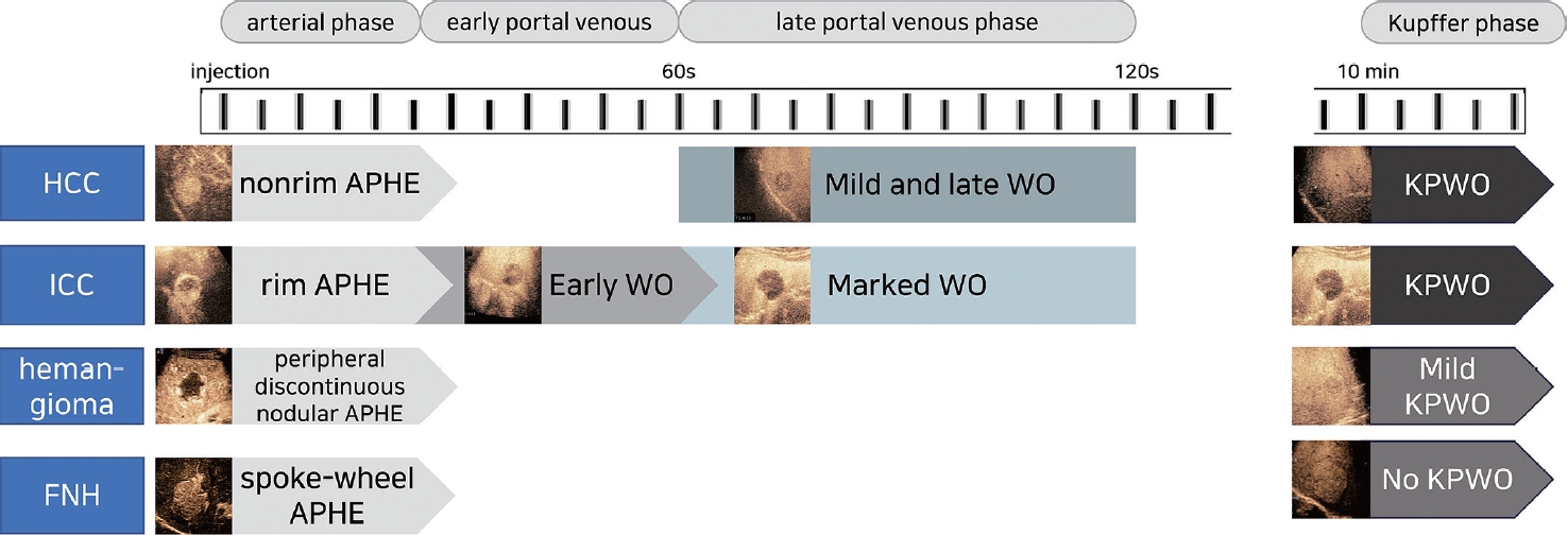
PDF - Sonazoid contrast-enhanced ultrasonography (CEUS) is a promising technique for the detection and diagnosis of focal liver lesions, particularly hepatocellular carcinoma (HCC). Recently, a collaborative effort between the Korean Society of Radiology and Korean Society of Abdominal Radiology resulted in the publication of guidelines for diagnosing HCC using Sonazoid CEUS. These guidelines propose specific criteria for identifying HCC based on the imaging characteristics observed during Sonazoid CEUS. The suggested diagnostic criteria include nonrim arterial phase hyperenhancement, and the presence of late and mild washout, or Kupffer phase washout under the premise that the early or marked washout should not occur during the portal venous phase. These criteria aim to improve the accuracy of HCC diagnosis using Sonazoid CEUS. This review offers a comprehensive overview of Sonazoid CEUS in the context of HCC diagnosis. It covers the fundamental principles of Sonazoid CEUS and its clinical applications, and introduces the recently published guidelines. By providing a summary of this emerging technique, this review contributes to a better understanding of the potential role of Sonazoid CEUS for diagnosing HCC.
-
Citations
Citations to this article as recorded by
- Sonazoid contrast-enhanced ultrasonography for the diagnosis of hepatocellular carcinoma: strengths and shortcomings
Sung Won Lee, Min Kyu Kang, Xiang Zhang
Journal of Liver Cancer.2023; 23(2): 238. CrossRef
- Sonazoid contrast-enhanced ultrasonography for the diagnosis of hepatocellular carcinoma: strengths and shortcomings

Original Article
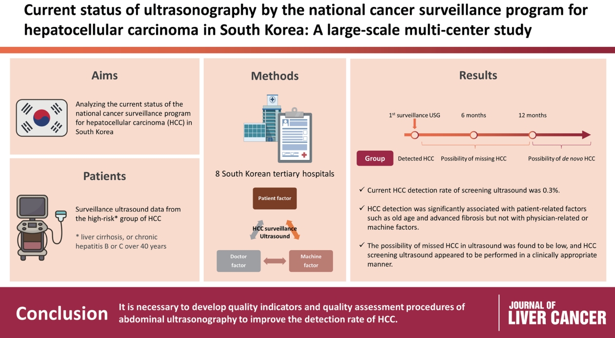
- Current status of ultrasonography in national cancer surveillance program for hepatocellular carcinoma in South Korea: a large-scale multicenter study
- Sun Hong Yoo, Soon Sun Kim, Sang Gyune Kim, Jung Hyun Kwon, Han-Ah Lee, Yeon Seok Seo, Young Kul Jung, Hyung Joon Yim, Do Seon Song, Seong Hee Kang, Moon Young Kim, Young-Hwan Ahn, Jieun Han, Young Seok Kim, Young Chang, Soung Won Jeong, Jae Young Jang, Jeong-Ju Yoo
- J Liver Cancer. 2023;23(1):189-201. Published online March 24, 2023
- DOI: https://doi.org/10.17998/jlc.2023.03.11
- 1,548 Views
- 65 Downloads
- 2 Citations
-
Abstract
PDF
 Supplementary Material
 - Background/Aim
Abdominal ultrasonography (USG) is recommended as a surveillance test for high-risk groups for hepatocellular carcinoma (HCC). This study aimed to analyze the current status of the national cancer surveillance program for HCC in South Korea and investigate the effects of patient-, physician-, and machine-related factors on HCC detection sensitivity.
Methods
This multicenter retrospective cohort study collected surveillance USG data from the high-risk group for HCC (liver cirrhosis or chronic hepatitis B or C >40 years of age) at eight South Korean tertiary hospitals in 2017.
Results
In 2017, 45 experienced hepatologists or radiologists performed 8,512 USG examinations. The physicians had a mean 15.0±8.3 years of experience; more hepatologists (61.4%) than radiologists (38.6%) participated. Each USG scan took a mean 12.2±3.4 minutes. The HCC detection rate by surveillance USG was 0.3% (n=23). Over 27 months of follow-up, an additional 135 patients (0.7%) developed new HCC. The patients were classified into three groups based on timing of HCC diagnosis since the 1st surveillance USG, and no significant intergroup difference in HCC characteristics was noted. HCC detection was significantly associated with patient-related factors, such as old age and advanced fibrosis, but not with physician- or machine-related factors.
Conclusions
This is the first study of the current status of USG as a surveillance method for HCC at tertiary hospitals in South Korea. It is necessary to develop quality indicators and quality assessment procedures for USG to improve the detection rate of HCC. -
Citations
Citations to this article as recorded by- The Epidemiology of Hepatitis B Virus Infection in Korea: 15-Year Analysis
Log Young Kim, Jeong-Ju Yoo, Young Chang, Hoongil Jo, Young Youn Cho, Sangheun Lee, Dong Hyeon Lee, Jae Young Jang
Journal of Korean Medical Science.2024;[Epub] CrossRef - Long-Term HBsAg Titer Kinetics with Entecavir/Tenofovir: Implications for Predicting Functional Cure and Low Levels
Soon Kyu Lee, Soon Woo Nam, Jeong Won Jang, Jung Hyun Kwon
Diagnostics.2024; 14(5): 495. CrossRef
- The Epidemiology of Hepatitis B Virus Infection in Korea: 15-Year Analysis
Review Article
- Contrast-enhanced Ultrasonography: The Third Modality for Differentiation of Liver Mass
- Min Kyu Kang, Moon Young Kim, Seong Hee Kang, Soon Koo Baik
- J Liver Cancer. 2019;19(2):91-96. Published online September 30, 2019
- DOI: https://doi.org/10.17998/jlc.19.2.91
- 4,644 Views
- 96 Downloads
- 1 Citation
-
Abstract
PDF
- Contrast-enhanced ultrasonography (CEUS) using microbubble ultrasonography contrast agent can show the vascular structure and unique contrast enhancement patterns of focal liver lesions, including hepatocellular carcinoma (HCC). CEUS shows three phases, similar to a vascular pattern on computer tomography (CT), and typical arterial enhancement and portal or late phase washout in HCC. CEUS can show real-time images without nephrotoxicity or radiation hazard and can be used as guidance for loco-regional treatment and estimation of treatment response of HCC. In addition, some data recently revealed the usefulness of CEUS in the early estimation of response to anti-cancer pharmacological (i.e., sorafenib) therapy in advanced HCC. Although CEUS has limitations in clinical practice and more investigation is needed for its validation, it is recommended as a main diagnostic modality in a few major clinical practice guidelines for HCC. Thus, greater understanding of CEUS is necessary to extend its application in real practice for diagnosis and management of diseases.
-
Citations
Citations to this article as recorded by- Perfluorobutane-Enhanced Ultrasound for Characterization of Hepatocellular Carcinoma From Non-hepatocellular Malignancies or Benignancy: Comparison of Imaging Acquisition Methods
Seungchul Han, Se Woo Kim, Sungeun Park, Jeong Hee Yoon, Hyo-Jin Kang, Jeongin Yoo, Ijin Joo, Jae Seok Bae, Jeong Min Lee
Ultrasound in Medicine & Biology.2023; 49(10): 2256. CrossRef
- Perfluorobutane-Enhanced Ultrasound for Characterization of Hepatocellular Carcinoma From Non-hepatocellular Malignancies or Benignancy: Comparison of Imaging Acquisition Methods
Original Article
- The Feasibility Study of Contrast-enhanced Ultrasound Using a 3 Dimension Transducer for Tumor Volume Measurement in Rabbit Hepatic VX2 Carcinoma Comparison with 2 Dimension Ultrasound and 3 Dimension Ultrasound without Contrast: Preliminary Results
- Kim, Jeehyun , Kim, Jung Hoon , Choi, Seo Youn , Han, Joon Koo
- J Liver Cancer. 2018;18(1):23-32. Published online March 31, 2018
- DOI: https://doi.org/10.17998/jlc.18.1.23
- 2,182 Views
- 24 Downloads
-
Abstract
PDF
- Background/Aim
s: To investigate the feasibility of tumor volume measurement using contrastenhanced ultrasound (US) with 3 dimension transducer (3D CEUS) in rabbit hepatic VX2 carcinoma.
Methods
Three different tumor volume measurements, including 2D US using the equation, 4/3(π)(abc), 3D US without contrast, and 3D CEUS were performed in 35 rabbit hepatic VX2 carcinomas. With the tumor volume from computerized tomography (CT) as a reference standard, we compared difference between CT volume and each different US tumor volume. The mean difference and correlation coefficient between each US volume measurement and CT volume were analyzed.
Results
Tumor volume measurement using 3D CEUS and 2D US using equation showed no statistical difference compared to CT volume (0.276 cm3, 0.212 cm3, and 0.263 cm3 vs. 0.306 cm3, 0.247 cm3, 0.276 cm3, P>0.05). However, 3D CEUS provided the highest correlation coefficient with CT volume (R=0.835 and 0.720) and the highest intraclass correlation (0.973 and 0.993). 3D CEUS provided a smaller mean difference with CT volume (0.016 cm3 and 0.033 cm3) than 2D US, showing 3D CEUS’s accurate measurement of tumor volume.
Conclusions
Due to its highly accurate, reliable, and reproducible measurements of tumor volume, 3D CEUS may be useful for predicting the therapeutic response evaluation after treatment.
Review Article
- Radiofrequency Ablation for Hepatocellular Carcinoma
- Se Young Jang, So Young Park
- J Liver Cancer. 2015;15(2):79-83. Published online September 30, 2015
- DOI: https://doi.org/10.17998/jlc.15.2.79
- 1,035 Views
- 18 Downloads
-
Abstract
PDF
- Radiofrequency ablation (RFA) takes an important role in management of hepatocellular carcinoma (HCC) as the most popular local therapy in the world. There are many data supporting that RFA is an excellent treatment modality for early-stage HCC with favorable treatment outcomes and minimal invasiveness. Currently, RFA extends treatement indications from unresectable early-stage HCC to intermediate-stage HCC in selected cases. Thus, with technical advances in guidance and ablation as well as devices, RFA widens its territory in the combat field against HCC. (J Liver Cancer 2015;15:79-83)
Original Article
- Follow-up of Hepatocellular Carcinoma After Transarterial Chemoembolization; The Concordance of Contrast Enhanced Ultrasonography and Lipiodol CT
- Gene Hyun Bok, Soung Won Jeong, Jae Young Jang, Sae Hwan Lee, Sang Gyune Kim, Sang-Woo Cha, Young Seok Kim, Young Deok Cho, Hong Soo Kim, Boo Sung Kim
- J Liver Cancer. 2014;14(2):115-119. Published online September 30, 2014
- DOI: https://doi.org/10.17998/jlc.14.2.115
- 882 Views
- 4 Downloads
-
Abstract
PDF
- Background/Aim
s: The aim of this study is to evaluate the concordance of contrast-enhanced ultrasonography (CEUS) and lipiodol computed tomography (L-CT) for the assessment of viable hepatocellular carcinoma (HCC) after transarterial chemoembolization (TACE).
Methods
We retrospectively reviewed the post-TACE CEUS and L-CT images of 65 consecutive HCCs in 41 patients to assess the presence of viable tumor tissue. Forty-seven HCCs in 31 patients that underwent post-TACE L-CT within 4 weeks of the CEUS examination were included. The degree of concordance between CEUS and L-CT and factors related to concordance were analyzed.
Results
The overall concordance of CEUS and LDCT was 78.7% (37/47). The concordance with L-CT for viable tumor and non-viable tumor tissue on CEUS was 95.2%, and 65.4% respectively (P<0.013). Diffuse tumors had a tendency for non-concordance (P=0.066). Although 3 of 4 lesions located in the hepatic dome were non-concordant, the sample size was too small to establish significance. The mean tumor size for concordant and non-concordant tumors was 2.9 and 3.0 cm, with no significant difference.
Conclusions
Although the concordance of CEUS and L-CT for viable tumor tissue was high, the concordance for non-viable tumor tissue was relatively low. Prospective studies using angiography as a gold standard should be performed in the future. (J Liver Cancer 2014;14:115-119)
Review Article
- The Usefulness of Contrast-Enhanced Ultrasonography in the Diagnosis of Hepatocellular Carcinoma
- Moon Young Kim
- Journal of the Korean Liver Cancer Study Group. 2014;14(1):7-13. Published online March 31, 2014
- DOI: https://doi.org/10.17998/jlc.14.1.7
- 980 Views
- 7 Downloads
-
Abstract
PDF
- Contrast enhanced ultrasonography (CEUS) using microbubble ultrasonography agent is able to show the vascular structure and enhancement patterns of lesions, so it has an worth in the diagnosis of hepatocellular carcinoma (HCC) which is a typical cancer that has a characteristic neovascularization. CEUS shows 3 phase vascular pattern like computer tomography (CT) typical arterial enhancement and portal or late wash out in HCC. CEUS can show a enhancement pattern of HCC in a real time and it has no nephrotoxicity or radiation hazard. Beyond the diagnosis, CEUS has also shown usefulness in the guidance of locoregional treatment and estimation of treatment response of HCC. In addition, recently, a few data which show a usefulness of CEUS in the early estimation of response after target therapy in the advanced HCC, also have been reported. However, CEUS has limitations in clinical practice yet and more wide investigation is needed for the validation of usefulness and wide application in clinical practice. However, CEUS also has many advantages in the field of the diagnosis and management of HCC, so in in this review, we are going to introduce CEUS and overview its clinical usefulness briefly.
 First
First Prev
Prev


 Follow JLC on Twitter