 E-submission
Search
- Page Path
- HOME > Search
Review Article
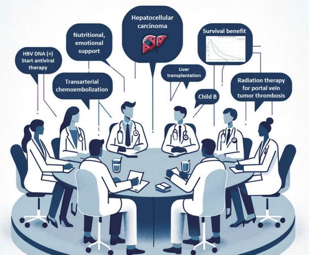
- Multidisciplinary approach for hepatocellular carcinoma patients: current evidence and future perspectives
- Joo Hyun Oh, Dong Hyun Sinn
- J Liver Cancer. 2024;24(1):47-56. Published online March 25, 2024
- DOI: https://doi.org/10.17998/jlc.2024.02.27
- 482 Views
- 44 Downloads
-
 Abstract
 PDF
PDF - Management of hepatocellular carcinoma (HCC) is challenging due to the complex relationship between underlying liver disease, tumor burden, and liver function. HCC is also notorious for its high recurrence rate even after curative treatment for early-stage tumor. Liver transplantation can substantially alter patient prognosis, but donor availability varies by each patient which further complicates treatment decision. Recent advancements in HCC treatments have introduced numerous potentially efficacious treatment modalities. However, high level evidence comparing the risks and benefits of these options is limited. In this complex situation, multidisciplinary approach or multidisciplinary team care has been suggested as a valuable strategy to help cope with escalating complexity in HCC management. Multidisciplinary approach involves collaboration among medical and health care professionals from various academic disciplines to provide comprehensive care. Although evidence suggests that multidisciplinary care can enhance outcomes of HCC patients, robust data from randomized controlled trials are currently lacking. Moreover, the implementation of a multidisciplinary approach necessitates increased medical resources compared to conventional cancer care. This review summarizes the current evidence on the role of multidisciplinary approach in HCC management and explores potential future directions.

Original Article
- Comparison of atezolizumab plus bevacizumab and lenvatinib for hepatocellular carcinoma with portal vein tumor thrombosis
- Jeayeon Park, Yun Bin Lee, Yunmi Ko, Youngsu Park, Hyunjae Shin, Moon Haeng Hur, Min Kyung Park, Dae-Won Lee, Eun Ju Cho, Kyung-Hun Lee, Jeong-Hoon Lee, Su Jong Yu, Tae-Yong Kim, Yoon Jun Kim, Tae-You Kim, Jung-Hwan Yoon
- J Liver Cancer. 2024;24(1):81-91. Published online January 19, 2024
- DOI: https://doi.org/10.17998/jlc.2023.12.25
- 982 Views
- 132 Downloads
-
Abstract
PDF
 Supplementary Material
 - Background/Aim
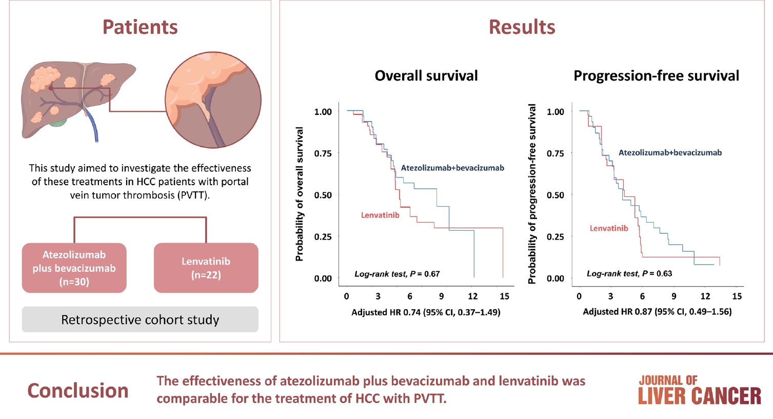
Atezolizumab plus bevacizumab and lenvatinib are currently available as first-line therapy for the treatment of unresectable hepatocellular carcinoma (HCC). However, comparative efficacy studies are still limited. This study aimed to investigate the effectiveness of these treatments in HCC patients with portal vein tumor thrombosis (PVTT).
Methods
We retrospectively included patients who received either atezolizumab plus bevacizumab or lenvatinib as first-line systemic therapy for HCC with PVTT. Primary endpoint was overall survival (OS), and secondary endpoints included progressionfree survival (PFS) and disease control rate (DCR) determined by response evaluation criteria in solid tumors, version 1.1.
Results
A total of 52 patients were included: 30 received atezolizumab plus bevacizumab and 22 received lenvatinib. The median follow-up duration was 6.4 months (interquartile range, 3.9-9.8). The median OS was 10.8 months (95% confidence interval [CI], 5.7 to not estimated) with atezolizumab plus bevacizumab and 5.8 months (95% CI, 4.8 to not estimated) with lenvatinib (P=0.26 by log-rank test). There was no statistically significant difference in OS (adjusted hazard ratio [aHR], 0.71; 95% CI, 0.34-1.49; P=0.37). The median PFS was similar (P=0.63 by log-rank test), with 4.1 months (95% CI, 3.3-7.7) for atezolizumab plus bevacizumab and 4.3 months (95% CI, 2.6-5.8) for lenvatinib (aHR, 0.93; 95% CI, 0.51-1.69; P=0.80). HRs were similar after inverse probability treatment weighting. The DCRs were 23.3% and 18.2% in patients receiving atezolizumab plus bevacizumab and lenvatinib, respectively (P=0.74).
Conclusion
The effectiveness of atezolizumab plus bevacizumab and lenvatinib was comparable for the treatment of HCC with PVTT.
Review Article
- Intrahepatic cholangiocarcinoma: histological diversity and the role of the pathologist
- Mina Komuta
- J Liver Cancer. 2024;24(1):17-22. Published online January 3, 2024
- DOI: https://doi.org/10.17998/jlc.2023.12.11
- 750 Views
- 95 Downloads
-
Abstract
PDF
- Intrahepatic cholangiocarcinoma (iCCA) is one of the primary liver cancers and presents with tumor heterogeneity. About 50% of iCCAs comprise actionable mutations, which completely change patient management. In addition, the precise diagnosis of iCCA, including subtype, has become crucial, and pathologists play an important role in this regard. This review focuses on iCCA heterogeneity; looking at different perspectives to guide diagnosis and optimal treatment choice.
Original Article
- The effects of immune checkpoint modulators on the clinical course of patients with resectable hepatocellular carcinoma
- Jihyun An, Hyo Jeong Kang, Eunsil Yu, Han Chu Lee, Ju Hyun Shim
- J Liver Cancer. 2022;22(1):40-50. Published online March 17, 2022
- DOI: https://doi.org/10.17998/jlc.2022.03.06

- 3,332 Views
- 111 Downloads
-
Abstract
PDFSupplementary Material
- Background/Aim
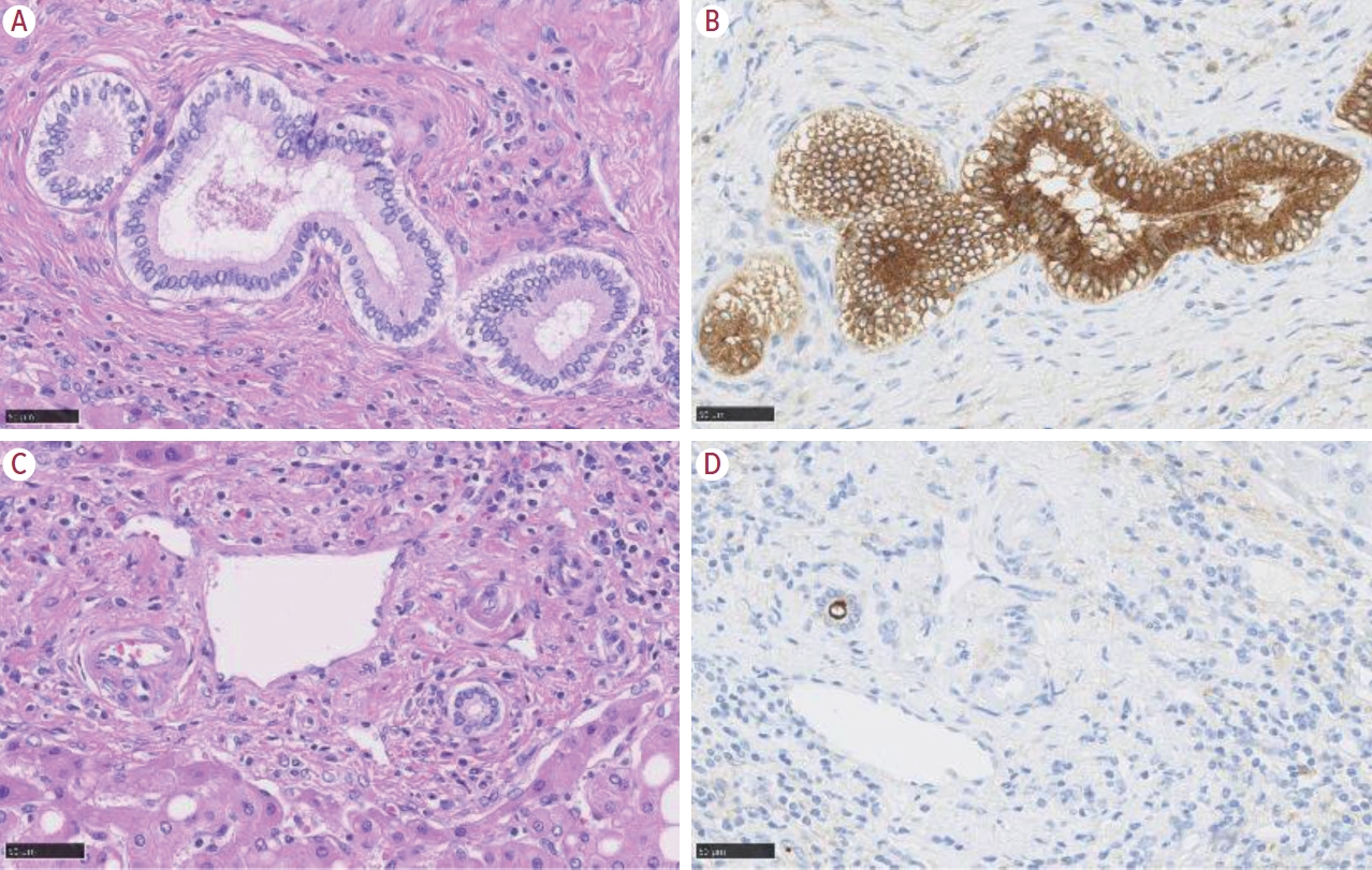
Immune checkpoint proteins regulating T-cell mediated anti-tumor immunity have been reported to affect clinical outcomes in multiple malignancies. This study aimed to investigate the prognostic effect of histological expression of immune checkpoint proteins in patients with resected hepatocellular carcinoma (HCC).
Methods
A total of 221 patients with HCC who underwent curative resection were included. Expression of programmed-cell death ligand-1 (PD-L1) in tumor cells (tPD-L1) and tumor infiltrating mononuclear cells (TIMCs) (iPD-L1), programmed-cell death-1 in TIMCs (iPD-1), and cytotoxic T lymphocyte antigen-4 in TIMCs (iCTLA-4) were measured immunohistochemically.
Results
Histo-positivity for iCTLA-4, iPD-1, iPD-L1, and tPD-L1 was 32.1%, 42.5%, 35.3%, and 14.9%, respectively. Multivariate logistic analyses revealed that male sex and tumor >5 cm were variables related to iCTLA-4 positivity (odds ratio [OR], 0.46 and 1.94, respectively; P<0.05). Poor differentiation was related to PD-L1 expression in both tumor cells and TIMCs (OR, 2.88 and 3.46, respectively; P<0.05). Microvascular invasion was significantly associated only with iPD-L1 (OR, 2.24; P<0.05). In time-dependent outcome analyses, expression of immune checkpoint proteins in TIMCs (i.e., iCTLA-4, iPD-1, and iPD-L1) was significantly related to longer overall survival and non-cancer-related survival (all P<0.05), but not to time-to-recurrence or cancer-specific deaths. Concurrent activation of the PD-1:PD-L1 and CTLA-4 pathways predicted improved outcomes in terms of overall survival and non-cancer related survival (P=0.06 and P=0.03, respectively).
Conclusions
Immune checkpoint proteins upregulated in TIMCs in HCC tissues have individual and additive effects in prolonging the survival of patients, specifically in terms of survival not related to cancer recurrence.
Review Article
- Differences in radiotherapy application according to regional disease characteristics of hepatocellular carcinoma
- Chai Hong Rim
- J Liver Cancer. 2021;21(2):113-123. Published online August 11, 2021
- DOI: https://doi.org/10.17998/jlc.2021.05.26
- 5,308 Views
- 101 Downloads
- 2 Citations
-
Abstract
PDF
- There are differences in opinion regarding the application of external beam radiotherapy in the treatment of hepatocellular carcinoma. Some major guidelines state that external beam radiotherapy is yet to attain a sufficient level of evidence. However, caution should be exercised when attempting to understand the clinical need for external beam radiotherapy solely based on the level of evidence. Previously, external beam radiotherapy had low applicability in the treatment of hepatocellular carcinoma before computed tomography-based planning was popularized. Modern external beam radiotherapy can selectively target tumor cells while sparing normal liver tissues. Recent technologies such as stereotactic body radiotherapy have enabled more precise treatment. The characteristics of hepatocellular carcinoma differ significantly according to the regional etiology. The main cause of hepatocellular carcinoma is the hepatitis B virus. It is commonly diagnosed as a locally advanced tumor but with relatively preserved hepatic function. The majority of these hepatocellular carcinoma cases are found in the East Asian population. Hepatocellular carcinoma caused by hepatitis C virus or other benign hepatitis tends to be diagnosed as a less locally aggressive tumor but with deteriorated liver function. The Western world and Japan tend to have patients with such causes. External beam radiotherapy has been more commonly performed for the former, although the use of external beam radiotherapy in the latter might have more concerns with regard to hepatic toxicity. This review discusses the above subjects along with perspectives regarding external beam radiotherapy in recent guidelines.
-
Citations
Citations to this article as recorded by
- Stereotactic Body Radiation Therapy for Hepatocellular Carcinoma: Meta-Analysis and International Stereotactic Radiosurgery Society Practice Guidelines
Sun Hyun Bae, Seok-Joo Chun, Joo-Hyun Chung, Eunji Kim, Jin-Kyu Kang, Won Il Jang, Ji Eun Moon, Isaure Roquette, Xavier Mirabel, Tomoki Kimura, Masayuki Ueno, Ting-Shi Su, Alison C. Tree, Matthias Guckenberger, Simon S. Lo, Marta Scorsetti, Ben J. Slotman
International Journal of Radiation Oncology*Biology*Physics.2024; 118(2): 337. CrossRef - Will the collaboration of surgery and external radiotherapy open new avenues for hepatocellular carcinoma with portal vein thrombosis?
Jung Wan Choe, Hye Yoon Lee, Chai Hong Rim
World Journal of Gastroenterology.2022; 28(7): 704. CrossRef
- Stereotactic Body Radiation Therapy for Hepatocellular Carcinoma: Meta-Analysis and International Stereotactic Radiosurgery Society Practice Guidelines
Case Report
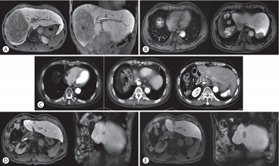
- A Case of Metastatic Melanoma in the Liver Mimicking Hepatocellular Carcinoma
- Jae-Kyoung So, Ji-Yun Hong, Min-Woo Chung, Sung-Bum Cho
- J Liver Cancer. 2021;21(1):92-96. Published online March 31, 2021
- DOI: https://doi.org/10.17998/jlc.21.1.92

- 6,922 Views
- 267 Downloads
-
Abstract
PDF
- The liver is one of the most common sites of metastasis. Although most metastatic liver cancers are hypovascular, some hypervascular metastases, such as those from melanoma, need to be differentiated from hepatocellular carcinoma (HCC) because they may show similar radiologic findings due to their hypervascularity. We encountered a case of multinodular liver masses with hyperenhancement during the arterial phase and washout during the portal venous and delayed phases, which were consistent with imaging hallmarks of HCC. The patient had a history of malignant melanoma and had undergone curative resection 11 years earlier. We performed a liver biopsy for pathologic confirmation, which revealed a metastatic melanoma of the liver. Metastatic liver cancer should be considered if a patient without chronic liver disease has a history of other primary malignancies, and caution should be exercised with hypervascular cancers that may mimic HCC.
Review Article
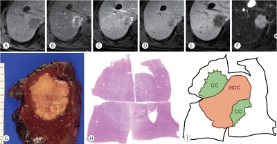
- Update on Pathologic and Radiologic Diagnosis of Combined Hepatocellular-Cholangiocarcinoma
- Hyungjin Rhee, Jae Hyon Park, Young Nyun Park
- J Liver Cancer. 2021;21(1):12-24. Published online March 31, 2021
- DOI: https://doi.org/10.17998/jlc.21.1.12
- 5,702 Views
- 298 Downloads
- 2 Citations
-
Abstract
PDF
- Combined hepatocellular-cholangiocarcinoma (cHCC-CCA) is a malignant primary liver carcinoma characterized by the unequivocal presence of both hepatocytic and cholangiocytic differentiation within the same tumor. Recent research has highlighted that cHCC-CCAs are more heterogeneous than previously expected. In the updated consensus terminology and WHO 2019 classification, “classical type” and “subtypes with stem-cell features” of the WHO 2010 classification are no longer recommended. Instead, it is recommended that the presence and percentages of various histopathologic components and stem-cell features be mentioned in the pathologic report. The new terminology and classification enable the exchange of clearer and more objective information about cHCC-CCAs, facilitating multi-center and multinational research. However, there are limitations to the diagnosis of cHCC-CCA by imaging and biopsy. cHCC-CCAs showing typical imaging findings of HCC could be misdiagnosed as HCC and subjected to inappropriate treatment, if other clinical findings are not sufficiently considered. cHCC-CCAs showing at least one of the CCA-like imaging features or unusual clinical features should be subjected to biopsy. There may be a sampling error for the biopsy diagnosis of cHCC-CCA. An optimized diagnostic algorithm integrating clinical, radiological, and histopathologic information of biopsy is required to resolve these diagnostic pitfalls.
-
Citations
Citations to this article as recorded by- Differentiation between hepatic angiomyolipoma and hepatocellular carcinoma in individuals who are not at-risk for hepatocellular carcinoma
Sungtae Park, Myeong-Jin Kim, Kyunghwa Han, Jae Hyon Park, Dai Hoon Han, Young Nyun Park, Jaehyo Kim, Hyungjin Rhee
European Journal of Radiology.2023; 166: 110957. CrossRef - The Human TOR Signaling Regulator Is the Key Indicator of Liver Cancer Patients’ Overall Survival: TIPRL/LC3/CD133/CD44 as Potential Biomarkers for Early Liver Cancers
Soo Young Jun, Hyang Ran Yoon, Ji-Yong Yoon, Su-Jin Jeon, Jeong-Ju Lee, Debasish Halder, Jin-Man Kim, Nam-Soon Kim
Cancers.2021; 13(12): 2925. CrossRef
- Differentiation between hepatic angiomyolipoma and hepatocellular carcinoma in individuals who are not at-risk for hepatocellular carcinoma
Case Reports
- Long-term Disease-free Survival after Trimodality Treatment of Recurrent Hepatocellular Carcinoma Involving the Inferior Vena Cava and Right Atrium
- Sunmin Park, Won Sup Yoon, Hyung Joon Yim, Chai Hong Rim
- J Liver Cancer. 2019;19(2):149-153. Published online September 30, 2019
- DOI: https://doi.org/10.17998/jlc.19.2.149
- 3,564 Views
- 73 Downloads
-
Abstract
PDFSupplementary Material
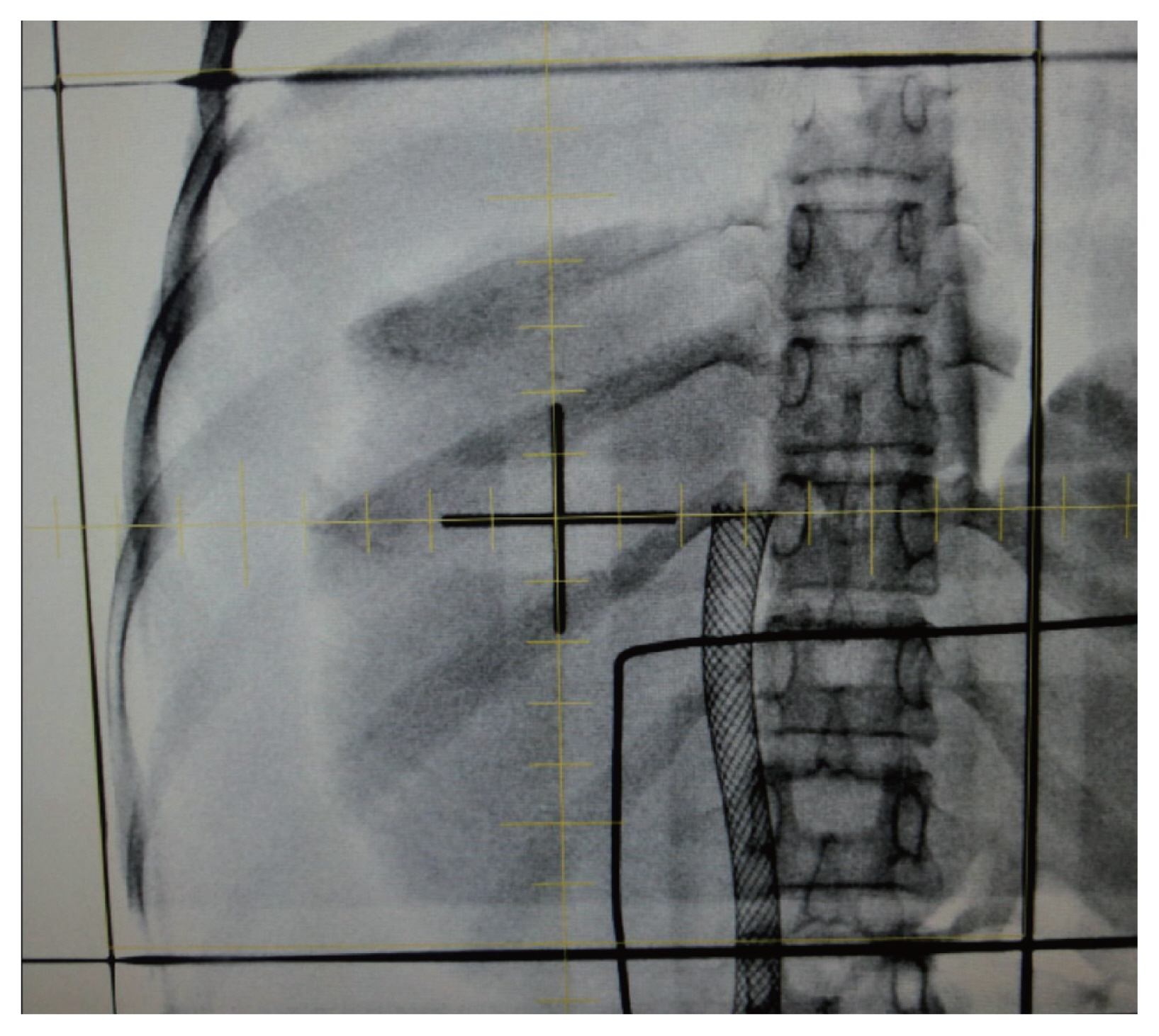
- Hepatocellular carcinoma (HCC) involving the inferior vena cava (IVC) and/or right atrium (RA) is a rare and intractable disease. A standard treatment has not been established yet, owing to the rarity of disease and difficulties in the therapeutic treatment. Herein, we report the case of a patient who had recurrent HCC (after a prior lobectomy) involving both IVC and RA and underwent multimodality treatments including external beam radiotherapy and transarterial chemotherapy, followed by sorafenib treatment. The disease was well controlled with local treatments and sustained for 7 years until last follow-up after the systemic treatments. Our case shows a possibility of long-term survival for patients affected by HCC involving IVC and/or RA, after a rigorous multimodality treatment strategy.
- Malignant Hepatic Solitary Fibrous Tumor
- Hong Il Kim, Seok Kyung In, Hyung Suk Yi, Min Jeong Lee, Hyo Young Kim
- J Liver Cancer. 2019;19(2):143-148. Published online September 30, 2019
- DOI: https://doi.org/10.17998/jlc.19.2.143

- 3,563 Views
- 51 Downloads
- 1 Citation
-
Abstract
PDF
- Hepatic solitary fibrous tumors (SFTs) are mostly benign and rare because of information regarding the clinical symptoms, treatment, and prognosis of their malignant forms is currently lacking. A literature review concerning malignant SFTs revealed that there were a few cases where patients experienced abdominal right upper quadrant (RUQ) pain as their first clinical symptom, and metastases were found after being diagnosed with hepatic SFT. Here, we report a patient who was previously healthy without any clinical symptoms such as RUQ pain or weight loss, but had the appearance of a metastatic mass as the first clinical presentation before a primary hepatic SFT was detected.
-
Citations
Citations to this article as recorded by- A Case of Hepatic Malignant Solitary Fibrous Tumor: A Case Report and Review of the Literature
Zhiyan Fu, Evita B. Henderson-Jackson, Barbara A. Centeno, Gregory Y. Lauwers, Mihaela Druta, Daniel A. Anaya, Yukihiro Nakanishi, Samir Sami Amr
Case Reports in Pathology.2023; 2023: 1. CrossRef
- A Case of Hepatic Malignant Solitary Fibrous Tumor: A Case Report and Review of the Literature
- Sarcomatoid Intrahepatic Cholangiocarcinoma: A Rare Case of Primary Liver Cancer
- Doo Hyuck Lee, Kyu Hyung Han, Sun Young Ahn, Sang Sun Kim, Hyun Sung Shin, Ki Bae Bang, Jun Ho Choi, Suk Bae Kim, Won Ae Lee, il Han Song
- J Liver Cancer. 2016;16(2):139-144. Published online September 30, 2016
- DOI: https://doi.org/10.17998/jlc.16.2.139
- 1,286 Views
- 17 Downloads
- 3 Citations
-
Abstract
PDF
- Sarcomatoid carcinoma arising from intrahepatic cholangiocyte, an extremely rare primary liver cancer, has highly invasive and metastatic potential. The pathogenesis of this tumor is unclear, although histogenetic mechanisms, such as transdifferentiation/dedifferentiation (epithelialmesenchymal transition or metaplastic transformation), biphasic differentiation (combination and collision), and redifferentiation, might be suggested to explain the simultaneous coexistence of carcinoma and sarcoma components in the same tumor. Immunohistochemical staining might be necessary to differentiate whether sarcomatous component is originated from hepatocyte or cholangiocyte. We report a case of sarcomatoid intrahepatic cholangiocarcinoma in a 58 year-old man presenting as an incidentally detected liver mass on regular health examination, which was diagnosed by an application of immunohistochemical methods after surgical resection, with a review of the literature based on 9 cases reported in Korea.
-
Citations
Citations to this article as recorded by- Pathologic features and clinical treatment of sarcomatoid intrahepatic cholangiocarcinoma
Xiaoli Xie, Nannan Lai, Yuanyuan Yang, Jinwei Zhang, Jianmin Qin, Xia Sheng
Intractable & Rare Diseases Research.2023; 12(4): 267. CrossRef - Clinical diagnosis and treatment strategies for sarcomatoid intrahepatic cholangiocarcinoma
Xia Sheng, Jian-Min Qin
World Chinese Journal of Digestology.2022; 30(14): 614. CrossRef - Analysis of intrahepatic sarcomatoid cholangiocarcinoma: Experience from 11 cases within 17 years
Dong Kyun Kim, Bo Ra Kim, Jin Sook Jeong, Yang Hyun Baek
World Journal of Gastroenterology.2019; 25(5): 608. CrossRef
- Pathologic features and clinical treatment of sarcomatoid intrahepatic cholangiocarcinoma
Original Article
- Academic Trend and Clinical Status of Radiotherapy for Hepatobiliary Cancer Over the Past 20 Years in Korea
- Won IL Jang, Yong-Seok Seo, Mi-Sook Kim, Heejin Kim
- J Liver Cancer. 2015;15(2):100-105. Published online September 30, 2015
- DOI: https://doi.org/10.17998/jlc.15.2.100
- 1,137 Views
- 18 Downloads
-
Abstract
PDF
- Background/Aim
s: To analyze the future trends through the status of radiotherapy in the hepatobiliary cancer in Korea and related articles published in the world.
Methods
Science citation index (SCI) and science citation index expanded (SCIE) articles, published in the 20 years from 1995 until 2014, were searched that contain the keywords related hepatobiliary cancer and radiotherapy using the Scopus. The incidence of hepatobiliary cancer was analyzed using annual reports from the Korea Central Cancer Registry. The status of radiotherapy was analyzed using data obtained form the Korean Society for Radiation Oncology and the National Health Insurance Service.
Results
Total 2,302 papers related radiotherapy for hepatobiliary cancer were searched in the world. By 2014, the cumulative number of papers published by domestic authors was a total 221 pieces. In 1999, total 16,305 hepatobiliary cancer patients were developed, of which 729 patients have been treated with radiotherapy. In 2013, it was expected that total 22,482 hepatobiliary cancer patients would be developed, of which 3,075 patients have been treated with radiotherapy.
Conclusions
Over the past 20 years, South Korea has made clinically and academically remarkable advances in the area of radiotherapy for hepatobiliary cancer. The researchers will continue to announce the results such as an objective status data and published papers in the future. (J Liver Cancer 2015;15:100-105)
Review Articles
- Clinical Safety and Efficacy of JX-594, a Targeted Multi-Mechanistic Oncolytic Pox Virus in Advanced Hepatocellular Carcinoma
- Jeong Heo
- Journal of the Korean Liver Cancer Study Group. 2011;11(2):149-154. Published online September 30, 2011
- 607 Views
- 4 Downloads
-
Abstract
PDF
- JX-594 is a targeted oncolytic vaccinia virus designed to selectively replicate in and destroy cancer cells with epidermal growth factor receptor (EGFR)/ras pathway activation. Direct oncolysis plus granulocyte macrophage-colony stimulating factor (GM-CSF) expression is accompanied by tumor vascular shutdown and anti-tumoral immunity. In a Phase 1 trial, JX-594 injection into hepatocellular carcinoma (HCC) was well-tolerated and associated with viral replication, anti-cancer immunity, decreased tumor perfusion and tumor necrosis. JX-594 has been shown to be well-tolerated by intravenous (IV) infusion and intratumoral (IT) injection and JX-594 is being developed as a novel therapy for patients with refractory or advanced HCC. Synergistic anti-tumor effects are predicted with JX-594 and sorafenib due to acute vascular shut down effect and tumor-specific antibody formation of JX-594 and chronic anti-angiogenic effects of sorafenib. JX-594 shows promise as a novel agent for the treatment of advanced HCC.
- Multistep Carcinogenesis of Hepatocellular Carcinoma
- Ja-June Jang
- Journal of the Korean Liver Cancer Study Group. 2008;8(1):39-46. Published online June 30, 2008
- 557 Views
- 17 Downloads
-
Abstract
PDF
- Epidemiological and experimental data have demonstrated that the process of carcinogenesis is progressive and multistage in nature. Model systems in animals exhibit this property of cancer development for several organ systems. The rat liver is one of the most extensively studied models of carcinogenesis. Multiple formats have been described for the analysis of cancer development in this organ, including the resistant hepatocyte selection regimens, the neonatal rat model and the partial hepatectomy model. The evolution of hepatic neoplasia is a slow process leading from the normal state via preneoplasia to benign and malignant neoplasia. On the histological level, hepatic preneoplasia usually emerges as foci of altered hepatocytes (FAH) which are perfectly integrated in the normal liver parenchyma and have no obvious neoplastic nature. The early emergence of FAH seems to be a general phenomenon of hepatocarcinogenesis in all species, no matter how this process has been elicited. The hallmark for the definition and detection of hepatic preneoplasia are biochemical and morphological changes in the hepatocellular phenotypes, which are neither uniform nor stable. In rodent liver treated with various chemical carcinogens, most of phenotypes have been shown to represent successive stages in an ordered sequence of cellular changes, progressing from glycogenic, clear and eosinophilic cell foci, through intermediate, mixed and basophilic cell populations, to hepatocellular adenomas and carcinomas, the fast growing variants of which consist of glycogen-poor, basophilic (ribosome-rich) cells. The identification of the placental isozyme of glutathione S-transferase (GST-P) as a highly expressed cytoplasmic protein during early carcinogenesis has led to its use as a marker of hepatic tumor development in early focal lesions, nodules and carcinomas. Different lesions have been suggested to represent preneoplastic conditions in human liver. They include large-cell change, small-cell change, foci of altered hepatocytes and dysplastic nodules. Experimental results suggest that multiple progressive factors are also involved in human hepatocarcinogenesis.
Case Reports
- Hepatoid Adenocarcinoma of the Stomach with Liver Metastasis Mimicking Hepatocellular Carcinoma
- Se Hyung Kim, Byung Ihn Choi
- Journal of the Korean Liver Cancer Study Group. 2003;3(1):65-68. Published online July 31, 2003
- 493 Views
- 5 Downloads
-
Abstract
PDF
- We describe a case of hepatoid adenocarcinoma of the stomach with liver metastasis mimicking hepatocellular carcinoma (HCC). Ultrasonography demonstrated an ill-defined heterogeneous hyperechoic mass at right lobe of the liver and high echoic thrombi that filled both portal veins. Contrast enhanced CT scan revealed an ill-defined low attenuated lesion in right lobe of the liver and left portal vein and posterior branch of right portal vein that were filled with low attenuated tumor thrombi. Diffuse and irregular wall thickening with enhancement was also visualized on the posterior wall of the stomach from cardia to angle. Endoscopic biopsy for gastric lesion and US-guided percutaneous core biopsy for hepatic mass were performed. Microscopic examination and immunohistochemical staining revealed aFP-producing hepatoid adenocarcinoma of the stomach and metastatic tumor of the liver.
- Undifferentiated Embryomal Sarcoma: A Case Report
- Jin Young Na, Won Jae Lee, Seung Hoon Kim, Hyo Keun Lim
- Journal of the Korean Liver Cancer Study Group. 2002;2(1):93-95. Published online July 31, 2002
- 5,756 Views
- 1 Download
-
Abstract
PDF
- A 60-year-old woman who admitted to our institute due to general malaise for 1 year was found to have a huge hepatic mass at ultrasound and CT. The hepatic tumor was histologically confirmed to be undifferentiated embryonal sarcoma by means of percutaneous needle biopsy under ultrasound guidance. Though this malignant hepatic tumor usually occurs in children and young adults, we report a case of undifferentiated embryonal sarcoma that occurred in an old woman.
 First
First Prev
Prev


 Follow JLC on Twitter