 E-submission
Articles
- Page Path
- HOME > J Liver Cancer > Volume 19(2); 2019 > Article
-
Case Report
Malignant Hepatic Solitary Fibrous Tumor -
Hong Il Kim1, Seok Kyung In1, Hyung Suk Yi1, Min Jeong Lee2, Hyo Young Kim1

-
Journal of Liver Cancer 2019;19(2):143-148.
DOI: https://doi.org/10.17998/jlc.19.2.143
Published online: September 30, 2019
1Department of Plastic and Reconstructive Surgery, Kosin University College of Medicine, Busan, Korea
2Department of Internal Medicine, Gupo Sungshim Hospital, Busan, Korea
- Corresponding author : Hyo Young Kim Department of Plastic and Reconstructive Surgery, Kosin University College of Medicine, 262 Gamcheon-ro, Seo-gu, Busan 49267, Korea tel. +82-51-990-6131, Fax. +82-51-990-6312 e-mail; hose3290@naver.com
Copyright © 2019 The Korean Liver Cancer Association
This is an Open Access article distributed under the terms of the Creative Commons Attribution Non-Commercial License (http://creativecommons.org/licenses/by-nc/4.0/) which permits unrestricted non-commercial use, distribution, and reproduction in any medium, provided the original work is properly cited.
- 3,554 Views
- 51 Downloads
- 1 Citation
Abstract
- Hepatic solitary fibrous tumors (SFTs) are mostly benign and rare because of information regarding the clinical symptoms, treatment, and prognosis of their malignant forms is currently lacking. A literature review concerning malignant SFTs revealed that there were a few cases where patients experienced abdominal right upper quadrant (RUQ) pain as their first clinical symptom, and metastases were found after being diagnosed with hepatic SFT. Here, we report a patient who was previously healthy without any clinical symptoms such as RUQ pain or weight loss, but had the appearance of a metastatic mass as the first clinical presentation before a primary hepatic SFT was detected.
- Solitary fibrous tumors (SFTs) are rare mesenchymal neoplasms usually found in the pleural cavity [1]. However, they can also originate in other body parts, including the retroperitoneum, trunk, extremities, head, and neck. SFT are mostly benign [2].
- Hepatic SFT is rare and is reported to be benign [3]. Only a few cases of malignant hepatic SFT have been reported, and right upper quadrant (RUQ) pain, weight loss, and loss of consciousness are some of the first symptoms to appear [4,5]. According to the available literature, the skull base, lung, and vertebrae showed metastases after malignant hepatic SFT was diagnosed. Here, we report a case of malignant hepatic SFT with metastatic subcutaneous mass as the first symptom in a previously healthy patient without abdominal or systemic symptoms, along with a literature review.
INTRODUCTION
- 1. Clinical findings
- In June 2017, a 69-year old woman was admitted to the hospital with a chief complaint of a mass in the right flank region. The patient was previously healthy without any past history, abdominal pain, anorexia, or weight loss findings. On physical examination, a 3×2 cm-sized, non-tender, oval-shaped mass was observed in the right flank subcutaneous region. Laboratory test results revealed elevated alkaline phosphatase (139 IU/L), gamma-glutamyltransferase (547 IU/L), aspartate aminotransferase (52 IU/L), and alanine aminotransferase (74 IU/L) levels, whereas carcinoembryonic antigen, carbohydrate antigen 19-9, and alpha-fetoprotein levels were within the normal range.
- 2. Diagnosis, image, and pathologic findings
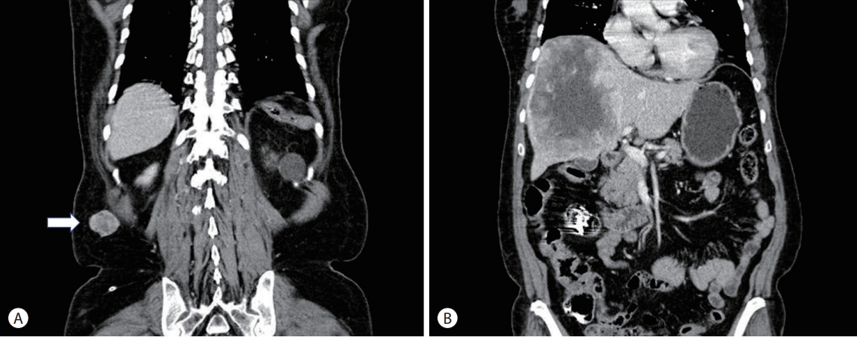
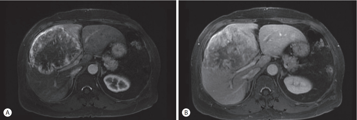
- Abdominal computed tomography (CT) revealed a mass in the right flank region with relatively clear margins (Fig. 1A), and its depth and size were evaluated. An incidental finding of a 14×11×10 cm-sized heterogeneous enhanced mass was also observed in the right hepatic lobe (Fig. 1B). The mass was evaluated using magnetic resonance imaging (MRI). Early arterial enhancement findings were noted in the liver third-phase MRI arterial phase (Fig. 2A), while delayed progress enhancement findings were observed in the portal phase (Fig. 2B), which was interpreted as a suspicious hypervascular sarcoma.
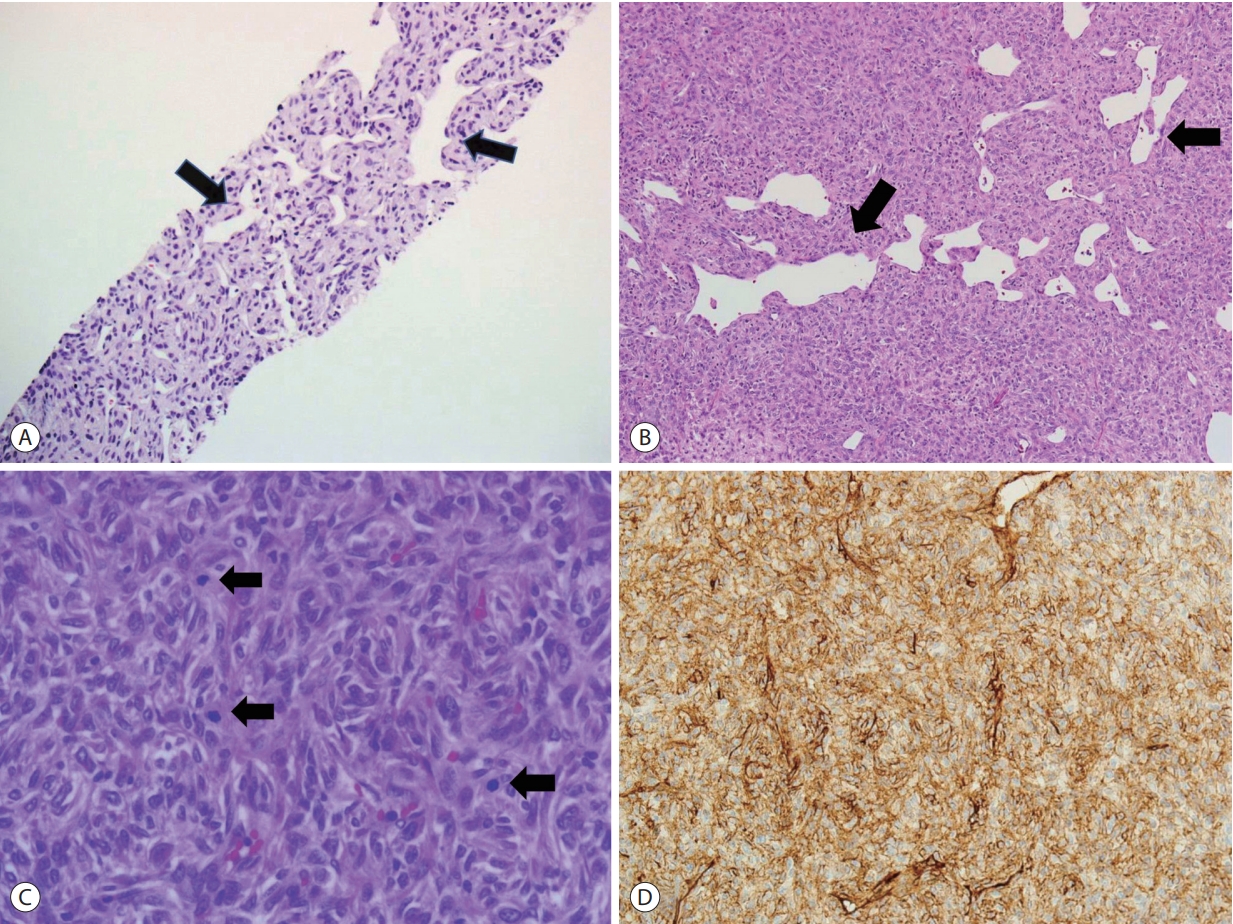
- Liver histological examination was performed using fine needle biopsy; the cells revealed similar morphology to that of spindled tumor cells with a typical “staghorn” vascular pattern of malignant SFT (Fig. 3A). For clinical staging, excisional biopsy of the flank mass was conducted; the histological analysis of the lesion was a “patternless” disposition with typical ectatic and irregularly shaped vessels (Fig. 3B). Additionally, in high power, tumor cells are ovoid to spindle shaped with relatively uniform and bland cytology, and increased mitotic activity was identified (Fig. 3C). The cluster of differentiation (CD) 34 immunohistochemical expression was strong and diffuse (Fig. 3D).
- 3. Treatment progress
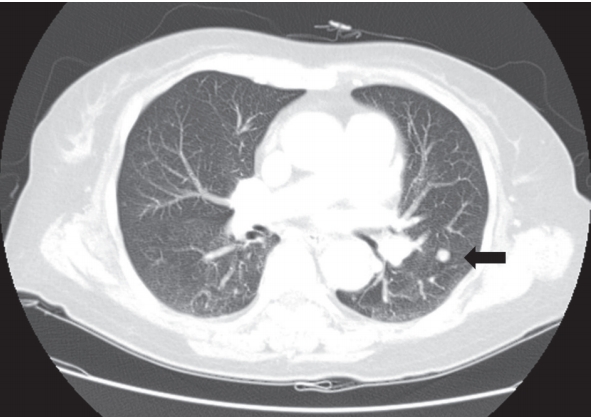
- Chest CT showed a suspected pulmonary metastasis (Fig. 4). The patient refused palliative surgery and thus, received three cycles of chemotherapy (CTx) that included adriamycin (30 mg on days 1-3), ifosfamide (2,355 mg on days 2-4), and mesna (1,256 mg IV 0 hours, 3 hours, and 6 hours after ifosfamide). Adjunctive radiation therapy was not administered. Despite CTx, the tumor rapidly enlarged and pulmonary metastases were detected with weight loss. The patient died in June, 2018.
CASE REPORT
- The average age for the occurrence of hepatic SFT is 50 years (16-84) and SFTs are more common in female than in male (2.2:1). Because of their rarity, SFTs have no established classification, underlying predisposing factors, incidence, treatment, and prognosis.
- Malignant hepatic SFTs are extremely rare and according to a literature review, only been five cases of malignant hepatic SFT have been reported [4-8]. Sex, age, first clinical symptom, size of mass, location, treatment, time to metastasis, and prognosis of reported malignant hepatic SFTs are summarized in Table 1.
- The average age of malignant hepatic SFT patients, including our case, was 50.6 years (24-69) and malignant hepatic SFT are more common in female than in male (2:1). The age range and sex distribution of malignant hepatic SFTs were similar to those of benign SFTs. Unlike benign SFTs, which frequently have no symptoms, malignant SFTs showed RUQ pain as its first symptom, and there were various cases with systemic symptoms, including weight loss and loss of consciousness. However, in our case, the patient had no abdominal or systemic symptoms and she was admitted to the hospital with a painless metastatic subcutaneous mass as the chief complaint. The size of benign SFTs varied (5-30 cm) and, occurred in both the left and right lobes [3], but the size of the malignant SFT was frequently larger (14-32 cm) and was mostly found in the right lobe.
- Resection was performed in most malignant SFT cases, and additional CTx was performed in some cases. In our case, when the patient was admitted, subcutaneous and lung metastases were already present; therefore, performing surgery was impossible. Thus, only CTx was administered. Many authors believe that postoperative adjuvant CTx and radiotherapy are helpful in unresectability or malignancy cases [6,7]. Currently, the combination of doxorubicin and ifosfamide is the standard systemic CTx regimen for many soft-tissue sarcoma subtypes. There was a report of a malignant SFT case in which CTx showed a response [8]. Archontaki et al. [7] and we used CTx, including adriamycin, ifosfamide, and mensa; however, there was no response. Additional investigations in a controlled, prospective trial on the use of CTx and its regimen to treat malignant SFTs are warranted. There was a report of using radiotherapy as an adjunctive therapy for head and neck SFTs to prevent recurrence [9]; however, no radiotherapy study has been conducted on malignant hepatic SFTs. Therefore, the effectiveness of radiation therapy for these tumors could not be assessed. Further studies are required to evaluate the effectiveness of radiotherapy as an adjunctive therapy for malignant hepatic SFTs.
- In previous reports, metastatic sites were the vertebrae, lung, and skull base. Our case was metastasized to the subcutaneous tissue unlike the previous case. The time to metastasis after malignant hepatic SFT diagnosis varied from immediately after diagnosis to 8 years. When the examining the subcutaneous mass, a hepatic mass was not suspected. This represents a rare case where the metastatic lesion was detected before hepatic SFT was diagnosed. The mechanism of how the tumor metastasizes from the liver to the subcutaneous tissue is unclear. Subcutaneous seeding has been reported after percutaneous liver injection. In this case, however, there was no iatrogenic procedures, including percutaneous injection, in which the subcutaneous metastasis was found before the primary liver legion. Furthermore, non-iatrogenic subcutaneous metastasis may occur. Therefore, an asymptomatic subcutaneous mass may be considered as the first symptom of malignant hepatic SFT.
- Usually, the prognosis of malignant SFT is poor. Yilmaz et al. [4] mentioned that by excision and CTx, including cyclophosphamide and adriamycin, patients could survive up to 6 months; however, there was no long term follow up. All reports published since then mention a poor prognosis.
- In hematological tests, benign SFTs usually show no abnormalities [3]. Including our case, other studies about malignant SFTs reported that tumor marker and liver function test results were non-specific and could not be used to test for the suspicion of malignant SFT.
- In radiological findings, SFT should be suspected if imaging reveals a solitary, highly vascular, well circumscribed, encapsulated mass that shows heterogeneous enhancement (owing to the varied density of the collagen component) in CT and MR images, especially if the enhancement is hypointense on the T2-weighted MRI and shows progressive enhancement in the delayed phase [10]. However, because it is difficult to distinguish malignant from benign tumors radiologically, immunochemical evaluation should be used for diagnosis [11].
- In our case, a similar morphology of spindled tumor cells with a typical “staghorn” vascular pattern was found in the liver histological findings. The tumor was well-circumscribed, with increased cellularity, a focal area of necrosis at low power, and increased mitotic activity at high power in the flank mass. Furthermore, CD34 immunohistochemical expression was strong and diffuse. Based on these characteristics, hepatocellular malignancy could be suspected. However, the CD34 antibody is not SFT specific and could be positive in both angiosarcoma and gastrointestinal stromal tumors [9]. Because malignant SFTs share many features with other neoplasms, can occur at practically any site, and have no specific immunohistochemical profile, they can be extremely difficult to distinguish from other spindle cell neoplasms [12]. The diagnosis of malignant SFT was made based on the typical “staghorn” vascular pattern observed in the liver biopsy and the increased mitosis cell cytology of the subcutaneous biopsy.
- In summary, malignant hepatic SFT is a rare neoplasm for which clinical symptoms, diagnosis, treatment, and prognosis are unclear. We report a case with a clinical symptom (painless metastatic subcutaneous mass) and metastatic course that are different from previous reports.
DISCUSSION
-
Conception and design: Hyo Young Kim
Acquisition of data: Seok kyung In
Data analysis and interpretation : Min Jeong Lee
Drafting the article : Hong Il Kim
Critical revision of the article: Hyung Suk Yi
Final approval of the version to be published: Hyo Young Kim
Article information
| Study | Year | Age (years) | Gender | Clinical presentation | Size (cm) | Lobe of liver | Treatment | Metastasis site | Time to metastasis | Follow up (months) | Prognosis |
|---|---|---|---|---|---|---|---|---|---|---|---|
| Yilmaz et al. [4] | 2000 | 25 | Female | Epigastric mass, weakness, anorexia, | 32×30×12 | Right | Excision+CTx | Vertebrae | 1 month after surgery | 6 | Alive |
| Jakob et al. [5] | 2007 | 70 | Male | RUQ pain, transient loss of consciousness | 27×24×12 | Right | Excision | Lung | 9 months after diagnosis | 12 | Died |
| Korkolis et al. [6] | 2010 | 54 | Male | No data | 17×12×8 | Right | Excision | Liver recurrence, skull base metastasis | 8 years after diagnosis | 96 | Died |
| Archontaki et al. [7] | 2011 | 24 | Female | RUQ pain, abdominal discomfort, | 30×17×15 | Right | Excision+CTx | Skull base | Initial diagnosis | 16 | Died |
| Park et al. [8] | 2013 | 62 | Female | RUQ pain, weight loss | 20×15×10 | Border on left lobe | Excision | No metastasis | No data | No data | No data |
| Our case | 2018 | 69 | Female | Metastatic mass of right flank | 14×11×10 | Right | CTx | Subcutaneous tissue, lung | Metastasis found first | 12 | Died |
- 1. Briselli M, Mark EJ, Dickersin GR. Solitary fibrous tumors of the pleura: eight new cases and review of 360 cases in the literature. Cancer 1981;47:2678−2689.ArticlePubMed
- 2. Hasegawa T, Matsuno Y, Shimoda T, Hasegawa F, Sano T, Hirohashi S. Extrathoracic solitary fibrous tumors: their histological variability and potentially aggressive behavior. Hum Pathol 1999;30:1464−1473.ArticlePubMed
- 3. Perini MV, Herman P, D’Albuquerque LA, Saad WA. Solitary fibrous tumor of the liver: report of a rare case and review of the literature. Int J Surg 2008;6:396−399.ArticlePubMed
- 4. Yilmaz S, Kirimlioglu V, Ertas E, Hilmioglu F, Yildirim B, Katz D, et al. Giant solitary fibrous tumor of the liver with metastasis to the skeletal system successfully treated with trisegmentectomy. Dig Dis Sci 2000;45:168−174.ArticlePubMed
- 5. Jakob M, Schneider M, Hoeller I, Laffer U, Kaderli R. Malignant solitary fibrous tumor involving the liver. World J Gastroenterol 2013;19:3354−3357.ArticlePubMedPMC
- 6. Korkolis DP, Apostolaki K, Aggeli C, Plataniotis G, Gontikakis E, Volanaki D, et al. Solitary fibrous tumor of the liver expressing CD34 and vimentin: a case report. World J Gastroenterol 2008;14:6261−6264.ArticlePubMedPMC
- 7. Archontaki M, Korkolis DP, Arnogiannaki N, Hatzijiannis S, Dendrinos P, Megapanos C, et al. Histologically malignant solitary fibrous tumour of the anterior thoracic wall: a case report and review of the literature. Case Rep Med 2010;2010:257167. ArticlePubMedPMCPDF
- 8. Park MS, Patel SR, Ludwig JA, Trent JC, Conrad CA, Lazar AJ, et al. Activity of temozolomide and bevacizumab in the treatment of locally advanced, recurrent, and metastatic hemangiopericytoma and malignant solitary fibrous tumor. Cancer 2011;117:4939−4947.ArticlePubMedPMC
- 9. Yang X, Zheng J, Ye W, Wang Y, Zhu HG, Wang LZ, et al. Malignant solitary fibrous tumors of the head and neck: a clinicopathological study of nine consecutive patients. Oral Oncol 2009;45:678−682.ArticlePubMed
- 10. Kandpal H, Sharma R, Gupta SD, Kumar A. Solitary fibrous tumour of the liver: a rare imaging diagnosis using MRI and diffusionweighted imaging. Br J Radiol 2008;81:e282−e286.ArticlePubMed
- 11. Fuksbrumer MS, Klimstra D, Panicek DM. Solitary fibrous tumor of the liver: imaging findings. Am J Roentgenol 2000;175:1683−1687.Article
- 12. Bishop JA, Rekhtman N, Chun J, Wakely Jr PE, Ali SZ. Malignant solitary fibrous tumor: cytopathologic findings and differential diagnosis. Cancer Cytopathol 2010;118:83−89.ArticlePubMed
References
Figure & Data
References
Citations
- A Case of Hepatic Malignant Solitary Fibrous Tumor: A Case Report and Review of the Literature
Zhiyan Fu, Evita B. Henderson-Jackson, Barbara A. Centeno, Gregory Y. Lauwers, Mihaela Druta, Daniel A. Anaya, Yukihiro Nakanishi, Samir Sami Amr
Case Reports in Pathology.2023; 2023: 1. CrossRef
 PubReader
 ePub Link
 Download Citation
 Follow JLC on Twitter